admin
TREATMENT WITH FOLK REMEDIES
Nowadays, more and more people suffer from a huge number of different diseases. These could be leg ailments or eye problems. Retinal dystrophy is one of the most common diseases that occurs in modern people. Fortunately, folk remedies can easily and inexpensively improve the condition of your eyes. The presented disease is a dystrophic transformation that forms in the macula of the eye. The reason for this color is a special pigment located in the central zone of the retina.
Characteristic features of detachment
Retinal detachment - treatment with folk remedies Source: LadySpecial.ru The retina is a thin tissue at the back of the eye that transmits visual impulses to the brain and allows you to interpret what you see.
A retinal detachment causes this layer to separate from the underlying tissue due to various reasons, such as trauma, bleeding, severe nearsightedness, medications, or surgery. Retinal detachment can lead to complete loss of vision, which usually requires urgent surgical correction. However, surgery may not completely restore vision, and retinal detachment may recur.
Therefore, you need to carefully study folk remedies that are effective for treating retinal detachment. The main symptom of retinal detachment is floating spots in front of the eyes.
Very often, when a retinal detachment occurs, a person notices that floating spots appear before the eyes, which become more noticeable if you look at them against a light background (especially against the sky!). These floaters are created from small clumps inside the jelly that forms the vitreous humor.
Whenever you move your eyeball, you will notice that these spots move; if you want to look directly at them, they will disappear from view. In most cases, eye floaters do not pose a serious threat to a person, but they can be very annoying.
If you notice a sudden increase in the number of floaters in front of your eyes, this may be a sign of damage to the internal structure of the eye. The vitreous is a jelly-like substance that is 98% water, but is about 4 times more viscous and acts as a shock absorber.
If the retina detaches or breaks appear in it, “sparks” or floating spots appear before the eyes. This disease is accompanied by loss of vision. See your doctor immediately as surgery may be required in this case.
Given the fact that most eye floaters eventually dissolve back into the fluid from which they originated, one way to combat them is to not treat them.
However, you can try a number of home remedies to get rid of floaters in front of your eyes. Antioxidants, vitamins A, E and C are known to help treat floaters.
Hemp oil contains a balanced amount of omega-3 fatty acids, making it one of the most effective folk remedies for treating eye floaters. Taurine is another antioxidant that helps dissolve any waste that accumulates in the eyes.
Ayurvedic treatment is also useful for treating retinal detachment. First, it can prevent the causes of retinal detachment by reducing swelling, bleeding, and damage to the inside of the eye due to medications or surgery.
Secondly, Ayurvedic remedies can be used to treat retinal detachment. In some cases, it is Indian folk remedies that can completely cure retinal detachment, especially in cases where surgery is not possible at all.
Ayurvedic remedies can correct the tissue damage in the eye and also eliminate all the causes responsible for this disease. Moreover, special and simple Panchkarma (Ayurvedic cleansing) procedures are designed to treat neurological and metabolic imbalances in the body.
This is important to eliminate the causes that led to retinal detachment. In addition to oral Ayurvedic remedies, applying medicated ghee to the eyes, drinking medicated ghee, and taking plain oil enemas are very effective.
In addition, for the treatment of retinal detachment, Ayurveda recommends the use of herbal remedies for a long time and in high doses, which have a specific effect on the eyes, retina, blood, as well as the walls of arteries and capillaries.
Traditional treatment for retinal detachment includes the use of a number of folk remedies that can prevent this disease and facilitate healing. Be sure to consult your doctor before using any of the following remedies.
The main role of folk remedies in the treatment of retinal detachment is the following effects:
- antihypertensive folk remedies reduce blood pressure in the eye;
- antidiabetic agents are used in cases of diabetes or prediabetes;
- Plants rich in antioxidants and vitamins are good for the retina.
Mistletoe is a very powerful antihypertensive agent that helps reduce intraocular pressure. Use an infusion of 1 tsp. raw materials for 1 cup of boiled water. If you have retinal detachment, use this remedy 1 cup 2 times a day.
In general, a combination of folk remedies and medical procedures can effectively treat retinal detachment. Only comprehensive treatment can eliminate the causes of this disease.
In this case, patients gradually regain full or almost full vision over several weeks or several months. Folk remedies “work” by providing a significant improvement in vision for patients suffering from retinal detachment.
The retina of the eye is a nervous tissue that has a complex and unique structure. Its main function is the perception of images, and with the help of photoreceptors, the conversion of a light impulse into a nerve impulse, which is then transmitted to the brain.
The blood vessels located under the retina provide its nutrition. Any damage to the retina of the eyes can cause deterioration in the functions of the visual apparatus.
Retinal detachment is one of the most common and serious pathologies of the visual organs, which requires the use of drastic measures, namely, urgent surgical intervention. In some cases, therapeutic treatment is prescribed.
With this pathology, the supply of oxygen and essential nutrients to the retina of the eyes is interrupted, which leads to serious visual impairment.
Recommendations for the treatment of diseases of the visual organs
During the first and second weeks, you need to brew three teaspoons of the mixture, taking into account that one hundred milligrams of water is used for one teaspoon of plants. For preparation, a container with a volume of half a liter is used.
- The broth should be cooked over low heat for ten minutes;
- after the broth is removed from the stove, it should be infused for twelve hours;
- then, once the broth has settled, it must be strained and a couple of tablespoons of honey added;
- It is recommended to consume the decoction one hundred grams before meals, warmed up;
- every two weeks, it is necessary to gradually increase the dosage of the collection.
To treat diseases of the organs of vision, and in particular retinal dystrophy, it is necessary to use the herbal mixture for three months.
To stop degenerative changes in the retina, you need to include in your diet more foods containing carotene, anthocyanosides, lutein, lycopene, and bioflavonoids.
Treatment of the retina with folk remedies cannot be abandoned halfway. Medicinal preparations do not act on a strictly defined area of the human body. Each of the ingredients has a beneficial effect on all processes occurring in the body. Consumption of the collection improves the condition of the vascular system, normalizes nutrient metabolism and strengthens the immune system. When the immune system is weakened, a person becomes susceptible to viruses and colds. As a result of disruption of the bronchi, an irritating cough appears. With a diagnosis such as retinal dystrophy, coughing very often causes tears in the retina.
Symptoms
The retina and choroid of the eyes function only together, therefore, if any of them is diseased, pathological processes can become irreversible. Patients may experience a variety of symptoms, but the diagnosis will be disappointing in each case.
Pathogenesis occurs quite quickly, which requires urgent intervention. There is one common symptom - the sharpness of perception of visual information decreases. Symptoms that may require treatment can be divided into several main stages:
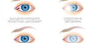
- Initial stage. Its main signs are photopsia, that is, the appearance of flashes, lightning and sparks in the eyes. They occur quite often, so the patient can easily identify them in himself. At the same time, failures in coordination of movements begin, and a sharp decrease in the perception of the clarity of objects is observed. Photopsia can appear in two cases: due to mechanical impact and in bright sunlight. A contraction occurs that stretches the retina of the eye, thus irritating the cells that have photosensitivity and creating these phenomena;
- Floating stage. This stage gets its name due to the appearance of characteristic symptoms. The patient may see floating threads, dots or floaters in the background of the image. These symptoms are not always an indicator of incipient retinal detachment. Sometimes this confirms the development of destruction of the vitreous body. But in any case, if such symptoms appear, you must definitely see a specialist for competent consultation and a full examination. You should not engage in self-treatment using folk remedies and methods;
- The final stage, which is characterized by the appearance of opacities. Experts call this effect the “Weiss ring.” It is this symptom that is a good reason to begin a more detailed study. At the final stage, not only retinal detachment can occur, but also detachment of the hyaloid posterior membrane. In this case, surgical treatment is mandatory and necessary.
The general condition that characterizes this process is expressed in the sudden appearance of several different symptoms at once: photopsia, opacities and loss of clarity of vision.
If the necessary therapeutic measures are not taken, the dissection may continue until hemorrhage occurs in the vitreous body, and then the process will be irreversible. The final stage, at which the final retinal detachment occurs, can threaten a person with complete loss of vision without the possibility of its restoration.
During hemorrhage into the vitreous body, blood vessels rupture, so even at the first signs of vision deterioration, diagnosis and appropriate treatment are required. The disease progresses very quickly.
At the very beginning of the development of such a pathology as retinal detachment, certain characteristic symptoms can be observed. If you do not pay attention to them, you may experience serious deterioration in health and even blindness.
Among the most basic symptoms that help resolve the question of how to determine retinal detachment pathology are:
- Decreased overall vision sharpness.
- Photopsia is the appearance of sparks and flashes before the eyes.
- Movement disturbance due to loss of sharpness of perception of nearby objects.
Such symptoms fully manifest themselves in two main situations - various methods of mechanical influence and bright sunlight. When faced with these factors, a strong contraction of the retina occurs, which in turn leads to irritation of receptor cells.
The patient begins to see spots, lightning and sparks before his eyes. A person who is experiencing retinal detachment may experience unpleasant flashes before their eyes. Already at this stage it is possible to judge with certainty that retinal detachment is developing.
Causes
The causes of this disease can be different. Timely hardware diagnostics and an experienced ophthalmologist who will take into account all the pathological processes occurring in the body that can affect vision will help identify the source.
Important
One of the causes of retinal detachment may be injury, which results in rupture of the membranes of the eye, including the retina.
If diseases affecting vision have previously been observed, then retinal detachment may result from untimely or ineffective treatment of the following pathologies:
- Damage to the vessels of the retina of the eyeballs in diabetes;
- Inflammation of the choroid;
- Eye tumors;
- Lesions of the central zone of the fundus.
Often healthy people may develop PVRD (peripheral vitreochorioretinal dystrophy), which causes a sharp deterioration in vision. Such pathologies are difficult to diagnose, so the doctor may not immediately understand the cause of vision problems.
In this case, a three-mirror Goldmann lens is required for a more thorough examination. There are a lot of risk factors, the main ones are:
- Various eye injuries;
- Visual defects with changes in the fundus;
- Difficult working conditions associated with exposure to high temperatures on the eye area;
- Genetic predisposition.
All patients who are at risk must be registered with an ophthalmologist and must undergo an annual examination and examination with a wide pupil.
Effect of E621 (monosodium glutamate) on the retina of the eye
The flavor enhancer E621, better known as monosodium glutamate, is well known to many consumers. It is often used by manufacturers of various food products and is especially popular due to the fact that it can improve and enhance the taste and smell of any food.
The synthetic food additive E621 is found in almost all products, the price of which is low and the quality is questionable. Japanese scientists, through numerous experiments and studies, have established the negative effect of E621 on the organs of vision.
In particular, it was noted that consumption of this synthetic food additive can cause the destruction of cells in the retina of the eyes as a result of the development of the process of apoptosis - programmed cell death and disruption of the structure of the retina, which in the future can become one of the causes of complete or partial loss of vision.
Only the synthetic form of monosodium glutamate has a similar effect. A natural flavor enhancer poses virtually no dangers.
Causes
Retinal dystrophy of the visual apparatus is a disease that affects older people. Age-related changes have a strong impact on the retina, causing it to become thinner. As the disease progresses, various tears and separations may appear in the affected areas over time.
Very often, when blood circulation is impaired, the process of nutrient metabolism deteriorates. As a result, various pathologies are observed in different structures of the eyeball.
The cause of the development of the disease may lie not only in age-related changes, but also be a complication of other serious diseases. Thus, impaired renal function, chronic hypertension, high myopia and diabetes mellitus can lead to serious vision problems. These diseases can be accompanied by loss of visual perception, attacks of dizziness, and in most cases lead to disruption of the heart muscle.
Treatment of the retina involves the use of folk remedies only as an auxiliary therapy
Treatment of retinal detachment
When pathology is identified, surgical intervention is often used. The effectiveness of treatment depends on the stage of the disease. Modern ophthalmology has very great capabilities.
The most progressive treatment methods are used, each of which is characterized by its own specific advantages and disadvantages, indications for use, which allows ophthalmologists to determine the most suitable option for each case.
Surgical techniques are considered the most effective and allow almost complete restoration of vision. These include:
- Cryopexy (freezing). With the help of special probes, the damaged retina is “glued together” and frozen at the rupture sites;
- Laser photocoagulation. This treatment uses powerful streams of light rays directed at the damaged area of the eye, resulting in microscopic burns around the retinal tears. After a burn, a scar will form to prevent the entry and accumulation of fluid;
- Vitreous removal or vitrectomy. This method allows the attending physician to gain free and most convenient access to the retina of the eye to eliminate the defect and cover existing extensive tissue tears;
- Sclerosis. In this case, in order to reduce pressure on the retina, prevent the formation of new tears and maintain the integrity of the layers of the retina, the attending physician places a fragment of elastic plastic, rubber or silicone in the area of the outer layer of the eye;
- Pneumatic retinopexy. An ophthalmologist injects an air bubble into the eye, which migrates to the area of the retinal tear and covers the damaged areas, preventing fluid from accumulating underneath them. Next, the doctor can use cryopexy (freezing) or photocoagulation (laser radiation) to connect the gap.
If you treat your health irresponsibly and do not consult an ophthalmologist in a timely manner at the first signs of retinal detachment, then the further development of the disease may result in:
- Hypotony of the eye;
- Development of cataracts;
- Subatrophy of the eyeball;
- Chronic iridocyclitis;
- Incurable blindness.
Before the operation, the patient undergoes a number of special diagnostic procedures and passes the necessary tests. The retina and fundus are examined, the main indicators are identified, visual acuity is checked, and a slit lamp examination is performed.
Since each case is individual, additional studies may be prescribed. Sometimes a chest x-ray and an ECG may be needed.
The patient must inform the attending physician about the presence of allergic reactions (if any) and about the constant use of any medications in order to avoid possible complications.
In this case, a week before the date of the scheduled operation, you must stop taking medications that thin the blood. 6 hours before the operation you should refuse food, unless there are contraindications, for example, with diabetes.
In this case, the doctor will advise you on how to prepare for the operation. During the operation, general or local anesthesia is used. The choice of anesthesia depends on the method of treatment, the general health of the patient, his age, weight, and the presence or absence of complications.
The duration of the procedure is about 2-4 hours. After the operation is completed, you may experience slight pain in the eye area and a feeling of nausea.
Most often, the patient can go home almost immediately, but it is better to agree in advance with relatives or loved ones about accompanying them, since weakness may occur.
For some time after the operation, you will need to follow the doctor's recommendations for a quick recovery. Immediately after all procedures, a sterile bandage is applied to the eye, which can only be removed with the permission of the attending physician, usually 24 to 36 hours after surgery.
For a month after surgery, the patient is not recommended to visit the sauna, and should also avoid rooms with high levels of humidity and high temperatures. During this period, it is necessary to ensure that water does not get into the eye and avoid heavy physical exertion.
You should strictly follow all the recommendations of your doctor and undergo a re-examination to avoid complications and speed up the process of restoring impaired visual functions.
Depending on the doctor’s instructions and compliance with all recommendations, you will be able to return to work within 1-2 weeks after the operation. Medication
Retinal detachment is accompanied by the death of photosensitive receptor cells that are responsible for visual acuity. Treatment with drugs will not work here, so you have to resort to surgery.
Surgical methods
The result of the operation depends on the stage at which it was performed. Currently, there are several options for surgical intervention, which are selected by a specialist depending on the situation.
The great advantage of operations is the possibility of complete restoration of vision. Surgical methods:
- Photocoagulation using laser. A stream of laser beams is directed into the eye. The rays cause micro burns in those places where retinal detachment occurred. Scars form at the site of burns, which prevent fluid from accumulating and getting under the retina.
- Cryopexy. This method is also called freezing. To perform the operation, probes are used that freeze the retina at the sites of detachment.
- Vitrectomy (removal of the vitreous body). To give the surgeon easy access to the retina, he may remove the vitreous. Thus, he will be able to eliminate damage to the retina.
- Pneumatic retinopexy. A small bubble of air is injected into the eye. It moves to the site of the retinal detachment and closes the tears and damage. This prevents liquid from accumulating there. To connect the detachment sites, the doctor can use laser photocoagulation or cryopexy.
- Sclerosis. For this purpose, a special elastic plastic or silicone is used, which is placed in the area of the outer layer of the eye. This method allows you to prevent further detachment and preserve the retina in its original state.
Surgical intervention (surgery)
Before resorting to surgical intervention, it is necessary to take tests (blood, urine, x-ray, electrocardiogram) and undergo diagnostics. You will need to check the condition of the retina and fundus of the eye. The ophthalmologist should check visual acuity and, if necessary, order additional tests.
Important
Before the operation, the doctor should find out about the presence of allergic reactions to medications. Before this, you should also not take blood thinning medications. Six hours before surgery you should not eat, but only if there are no contraindications.
During surgery. The patient is injected with anesthesia, which can be either local or general. It all depends on the method of surgery and the characteristics of the health condition. The operation lasts from two to four hours. When the patient wakes up after surgery, he will feel slight pain and nausea. In most cases, he can go home almost immediately.
After operation. To recover, you must follow all the specialist’s recommendations. After the operation, a bandage is put on the eye, which cannot be removed until the doctor’s permission. Most often, the bandage is removed a day or a day and a half after the operation.
For the next month after the operation, you should not go to the bathhouse or sauna, and try not to visit places with high humidity and temperature. Avoid getting water in your eyes, lifting heavy objects, and engaging in strenuous exercise.
To monitor the recovery process or, if problems arise, to detect them in a timely manner, you must visit an ophthalmologist and follow all his recommendations.
Possible complications
In rare cases, complications may occur after surgery. They are usually associated with cataracts, glaucoma, and the general poor health of the patient. The most common complications include:
- Recurrent retinal detachment, which will require new surgical intervention;
- Increased scarring of the retina (ie, proliferative vitreoretinopathy). In this case, repeated operational measures are also carried out;
- The development of endophthalmitis as a result of infection in the eye.
You should immediately consult a doctor if there is discharge from the eyes, fever, chills, swelling and redness, shortness of breath, cough, or pain in the chest area.
Treatment of retinal detachment with folk remedies - is it effective?
https://youtu.be/4gYkYElqMO8
Retinal detachment is one of the most serious diseases of the visual system, which can lead to complete loss of vision. Unfortunately, the mistrust or doubts of some patients regarding the capabilities of official medicine often leads them to search for alternative treatment methods.
Therefore, in the ophthalmologist’s office you can often hear the question: are there folk remedies and methods for treating retinal detachment? In order to answer this question correctly and as clearly as possible, it is necessary to clearly explain the mechanism of retinal detachment.
At its core, retinal detachment is a separation of the retina from its underlying tissue, mechanical damage, an open wound that cannot be treated in any way other than surgery.
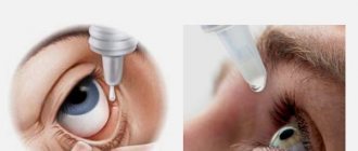
The only difference in surgical techniques lies solely in their complexity and the final results. Even the most miraculous compresses, vitamin drops, aloe juice, decoctions and other remedies available in the arsenal of traditional medicine will not allow the damaged retina to “grow back” to its original place.
Special gymnastics and eye exercises, which are presented by various healers as time-tested oriental techniques, will also be powerless. Any assurances of the benefits of traditional methods of treating retinal detachment and beliefs in their effectiveness are fundamentally incorrect and criminal.
Minor improvements when using such alternative methods may be observed only in the early stages of the disease. But this effect is temporary, which can only be explained by a sharp activation and mobilization of the regenerative reserves of the eyes, but this does not eliminate the main cause - the physical detachment of the retina.
Even officially approved, certified and most expensive eye drops cannot replace the effectiveness of surgery to restore a detached retina. Sooner or later, the patient will still have to see an ophthalmologist for surgical measures.
And it’s better if it’s early than late, when the disease reaches an advanced stage. Therefore, at the first symptoms, you must immediately consult a doctor. Any delay caused by experiments with traditional medicine and alternative treatments only greatly increases the risks of complete loss of vision.
Treatment with folk remedies
Source: tvoiglazki.ru Retinal detachment is a dangerous pathology that can lead to complete blindness. Many people do not understand the seriousness of the problem and are in no hurry to see a doctor, believing that traditional medicine will be more effective in treating detachment.
Retinal detachment occurs when the retina is torn from the underlying tissue due to some mechanical injury, trauma, or injury that can only be treated through surgery.
Therefore, no folk remedies or eye exercises can make the retina grow back again. Treatment with folk remedies can have a beneficial effect, but only at an early stage.
Unfortunately, the improvement will not last long, and the cause of the detachment itself will not be eliminated. Not all medications, such as eye drops, can help in this situation. Therefore, a person will still need to see a doctor, otherwise complications cannot be avoided.
Important
Retinal detachment is a fairly dangerous pathology that can lead to blindness if left untreated. The majority of people, when the first signs appear, are in no hurry to get help from a specialist, not attaching much seriousness to this disease.
It is simply impossible to cure detachment completely without surgery. Traditional methods of treatment can only help in the process of rehabilitation after surgery, as well as at the very initial stage, when detachment has not yet occurred, but the doctor has established the presence of an incipient problem.
Properly selected treatment methods can significantly strengthen the retina and prevent the development of a more complex form of pathology and its unpleasant consequences. When answering the question of how to treat retinal detachment at home, you can pay attention to certain effective methods of alternative treatment.
Spices for retinal diseases
Treatment of retinal dystrophy with folk remedies includes the ingestion of spices. It is important to remember that when choosing any ingredient you need to pay attention to contraindications. For example, marjoram is not recommended for use if there is a tendency to increased blood clotting, and cumin is not recommended for thrombophlebitis and cholelithiasis. Spices such as:
- dill;
- coriander;
- caraway;
- ginger root;
- laurel;
- cinnamon;
- carnation;
- garlic.